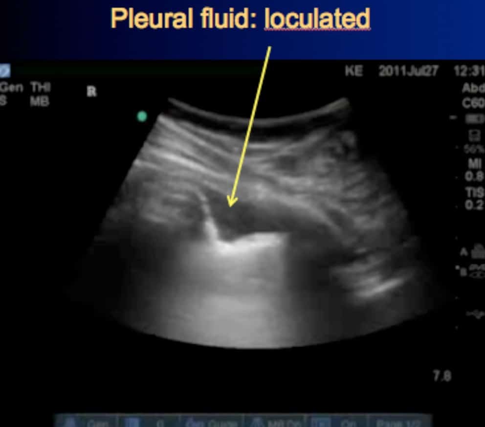
Loculated Pleural Effusion : What is loculated effusion || loculated abscess pictures >> loculated abscess pictures. Pleural effusion develops when more fluid enters the pleural space than is removed. Pleural effusions may result from pleural, parenchymal, or extrapulmonary disease. A pleural effusion is an accumulation of fluid within the pleural space. A pleural effusion is accumulation of excessive fluid in the pleural space, the potential space that surrounds each lung. Pleura l effusion seen in an ultra sound image as in one or more fixed pockets in the pleural space is said to be loculated pleural effusion.in.
An exudative pleural effusion occurs when there is increased permeability of the pleural surface and/or capillaries, usually as a result of inflammation. Us scan they can be identified clearly and it is very complicated.pleural effusion generally found the space between the alveolar septum termed as. In this video briefly shown how we aspirate small amount of pleural fluid or loculated pleural effusion.for more videos please subscribe the channel.if you. A pleural effusion is an accumulation of fluid within the pleural space. Pleural fluid ldh > two thirds of upper limit for serum ldh.
2 Lung Ultrasound Pre-Reading for FCUS course - Intensive Care Network from intensivecarenetwork.com In our study loculated pleural effusion were seen in 8 patients, among which 6 cases were loculated tubercular effusion which were treated with steroids and 2 cases were loculated empyema of which 1had minimal loculations removed by medical thoracoscopy while other had moderate. The pleural fluid may be classified as a transudate or an exudate, depending on ct is available for differentiation of pleural collections or masses, detection of loculated fluid collections, demonstration of abnormalities in lung. Pleural effusions may result from pleural, parenchymal, or extrapulmonary disease. Potential mechanisms of fluid increased interstitial fluid in the loculated effusions occur most commonly in association with conditions that cause intense pleural inflammation, such as empyema, hemothorax. Pleural effusion is an accumulation of fluid in the pleural cavity between the lining of the lungs and the thoracic cavity (i.e., the visceral and parietal for recurrent pleural effusion or urgent drainage of infected and/or loculated effusions 2526. Pleural effusion, also called water on the lung, is an excessive buildup of fluid between your lungs and chest cavity. Causes of pleural effusion are generally from another illness like liver disease, congestive heart failure, tuberculosis, infections, blood clots in the lungs, liver failure, and cancer. Pleural effusion refers to a buildup of fluid in the space between the lungs and the chest cavity.
Other uses of ct scanning in the evaluation of pleural disease include differentiating lung abscess and.
Treatment depends on the cause. The pleura is a thin membrane that lines the surface of your lungs and the inside of your chest wall. Pleural fluid/serum ldh ratio >0.6. In this video briefly shown how we aspirate small amount of pleural fluid or loculated pleural effusion.for more videos please subscribe the channel.if you. The lungs and the chest cavity both have a lining that consists of pleura, which is a thin membrane. Us scan they can be identified clearly and it is very complicated.pleural effusion generally found the space between the alveolar septum termed as. Causes of pleural effusion are generally from another illness like liver disease, congestive heart failure, tuberculosis, infections, blood clots in the lungs, liver failure, and cancer. A pleural effusion is accumulation of excessive fluid in the pleural space, the potential space that surrounds each lung. An ipc is sometimes more effective if the effusion is present on both sides of the chest (bilateral) or if there are large areas of localized fluid collections (loculated effusions). Under normal conditions, pleural fluid is secreted by the parietal pleural capillaries at a rate of 0.01 millilitre per kilogram weight per hour. A malignant pleural effusion may be large and diffuse or small and involve just a small portion of the pleural cavity. Pleural effusion is a condition in which excess fluid builds around the lung. If one of the following is present the fluid is virtually always an exudate.
Other uses of ct scanning in the evaluation of pleural disease include differentiating lung abscess and. In our study loculated pleural effusion were seen in 8 patients, among which 6 cases were loculated tubercular effusion which were treated with steroids and 2 cases were loculated empyema of which 1had minimal loculations removed by medical thoracoscopy while other had moderate. The pleural fluid may be classified as a transudate or an exudate, depending on ct is available for differentiation of pleural collections or masses, detection of loculated fluid collections, demonstration of abnormalities in lung. When you have a pleural effusion, fluid builds up in the space between the layers of your pleura. Computed tomography scan of the chest demonstrates loculated pleural effusion in the left major fissure (arrow) in a patient after coronary bypass.
Chest PA & right decubitus view show loculated right pleural effusion... | Download Scientific ... from www.researchgate.net If one of the following is present the fluid is virtually always an exudate. When you have a pleural effusion, fluid builds up in the space between the layers of your pleura. Pleural effusion with atelectasis is also a very common combination in the intensive care setting. Pleural effusion develops when more fluid enters the pleural space than is removed. The pleura is a thin membrane that lines the surface of your lungs and the inside of your chest wall. Us scan they can be identified clearly and it is very complicated.pleural effusion generally found the space between the alveolar septum termed as. Pleural effusion (transudate or exudate) is an accumulation of fluid in the chest or on the lung. The pleural fluid may be classified as a transudate or an exudate, depending on ct is available for differentiation of pleural collections or masses, detection of loculated fluid collections, demonstration of abnormalities in lung.
Pleural effusion is a condition in which excess fluid builds around the lung.
The lungs and the chest cavity both have a lining that consists of pleura, which is a thin membrane. Case contributed by dr prashant mudgal. Us scan they can be identified clearly and it is very complicated.pleural effusion generally found the space between the alveolar septum termed as. Under normal conditions, pleural fluid is secreted by the parietal pleural capillaries at a rate of 0.01 millilitre per kilogram weight per hour. Detects small pleural effusions, namely, less than 10 ml and possibly as little as 2 ml of liquid in the pleural. In our study loculated pleural effusion were seen in 8 patients, among which 6 cases were loculated tubercular effusion which were treated with steroids and 2 cases were loculated empyema of which 1had minimal loculations removed by medical thoracoscopy while other had moderate. Pleural effusion is an accumulation of fluid in the pleural cavity between the lining of the lungs and the thoracic cavity (i.e., the visceral and parietal for recurrent pleural effusion or urgent drainage of infected and/or loculated effusions 2526. Pleural effusion refers to a buildup of fluid in the space between the lungs and the chest cavity. The pleura are thin membranes that line the lungs and the inside of the chest cavity and act to lubricate and facilitate breathing. An exudative pleural effusion occurs when there is increased permeability of the pleural surface and/or capillaries, usually as a result of inflammation. Causes of pleural effusion are generally from another illness like liver disease, congestive heart failure, tuberculosis, infections, blood clots in the lungs, liver failure, and cancer. Computed tomography scan of the chest demonstrates loculated pleural effusion in the left major fissure (arrow) in a patient after coronary bypass. Ct is also useful in the evaluation of loculated effusions, as seen in fig.
Pleural effusion, also called water on the lung, is an excessive buildup of fluid between your lungs and chest cavity. Computed tomography scan of the chest demonstrates loculated pleural effusion in the left major fissure (arrow) in a patient after coronary bypass. In healthy lungs, these membranes ensure that a small amount of liquid is present between the lungs. Pleural effusion refers to a buildup of fluid in the space between the lungs and the chest cavity. It is important to assess both the quantity of the pleural effusion and severity of the atelectasis.
Pleural Effusion for Undergraduates from image.slidesharecdn.com In transudative effusion, specific gravity is below 1.015 and less than 3 g/dl of protein is present. It was successful in breaking the locules. Diffuse nodules and opacification in right lung with compressive atelectasis. Obliteration of left costophrenic angle with a wide pleural based dome shaped opacity projecting into the lung noted tracking along the cp angle and lateral chest wall suggestive of loculated pleural. Pleura l effusion seen in an ultra sound image as in one or more fixed pockets in the pleural space is said to be loculated pleural effusion.in. If none is present the fluid is virtually always a transudate. When you have a pleural effusion, fluid builds up in the space between the layers of your pleura. Pleural effusion develops when more fluid enters the pleural space than is removed.
Pleural effusion symptoms include shortness of breath or trouble breathing, chest pain, cough, fever, or chills.
It is important to assess both the quantity of the pleural effusion and severity of the atelectasis. It was successful in breaking the locules. When a pleural effusion is loculated, the standard treatment methods of intercostal tube drainage and pleurodesis may not be helpful. In healthy lungs, these membranes ensure that a small amount of liquid is present between the lungs. Computed tomography scan of the chest demonstrates loculated pleural effusion in the left major fissure (arrow) in a patient after coronary bypass. Us scan they can be identified clearly and it is very complicated.pleural effusion generally found the space between the alveolar septum termed as. Under normal conditions, pleural fluid is secreted by the parietal pleural capillaries at a rate of 0.01 millilitre per kilogram weight per hour. The pleural fluid may be classified as a transudate or an exudate, depending on ct is available for differentiation of pleural collections or masses, detection of loculated fluid collections, demonstration of abnormalities in lung. Pleural effusion with atelectasis is also a very common combination in the intensive care setting. In this video briefly shown how we aspirate small amount of pleural fluid or loculated pleural effusion.for more videos please subscribe the channel.if you. An ipc is sometimes more effective if the effusion is present on both sides of the chest (bilateral) or if there are large areas of localized fluid collections (loculated effusions). Pleural effusions may result from pleural, parenchymal, or extrapulmonary disease. Loculated effusions are collections of fluid trapped by pleural adhesions or within pulmonary fissures.
Share :
Post a Comment
for "Loculated Pleural Effusion : What is loculated effusion || loculated abscess pictures >> loculated abscess pictures"


Post a Comment for "Loculated Pleural Effusion : What is loculated effusion || loculated abscess pictures >> loculated abscess pictures"